Ultrafast Electron Crystallography
The emergence of ultrafast electron diffraction techniques has enabled the study of structural dynamics with both spatial and temporal resolution at the atomic level (~0.1 nm and ~100 fs). These techniques can provide valuable new insights in materials science and the life sciences. Examples are real-time observation of coherent phonons in solid-state physics and monitoring conformational changes in proteins, which ultimately determine the biological functions of these complex macromolecules. X-ray free electron lasers are able to produce X-ray pulses of sufficient brightness to perform single-shot, femtosecond diffraction on complex crystals, but also require large and expensive facilities. Ultrafast electron diffraction setups offer a compact and affordable alternative. In fact, ultrafast electron and X-ray diffraction techniques provide complementary information. By now, femtosecond electron diffraction has been applied successfully to study ultrafast (reversible) dynamics in gases and thin films [1].
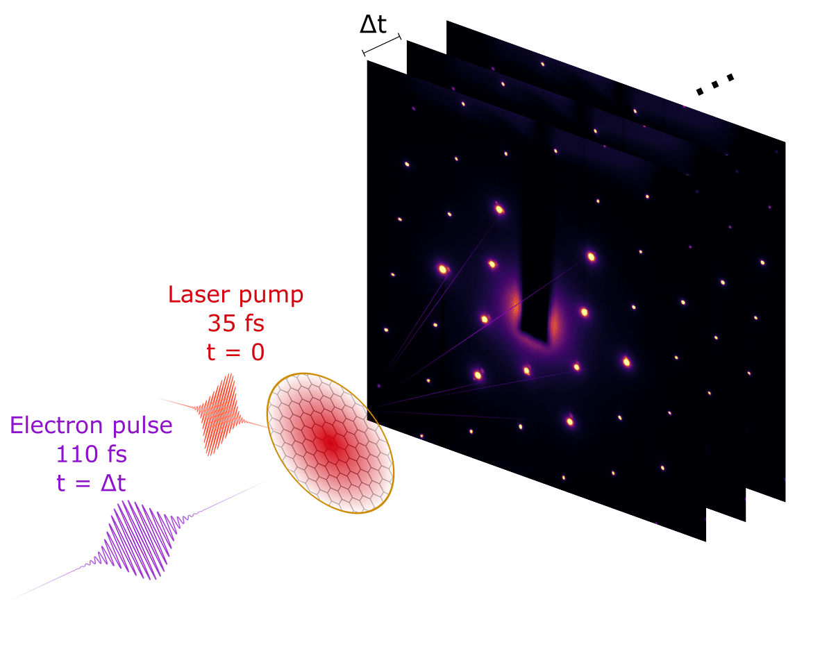
To enable systematic studies of dynamic systems using ultrafast electron diffraction (UED), a setup has been designed and developed by DrX Works for single-shot UED with electron bunches of 100 keV, 100 μm, 100 fs, and 100 fC, based on femtosecond photoemission and bunch compression with a radio-frequency (RF) cavity. Only ~100 fC, corresponding to roughly one million electrons, is required for recording a single diffraction pattern, but when confined in a single bunch of only 100 fs duration, this inevitably leads to a strong space-charge explosion and thus a rapid increase of the bunch duration. By using a 3 GHz RF cavity in TM-010 mode, which is synchronized to the femtosecond photoemission laser, this bunch explosion can be reversed and the original 100 fs bunch duration restored. This technique has proven to be successful and has been adopted by many research groups all over the world. The video below explains the operation of the photogun and the compression cavity in an ultrafast diffraction experiment.
Single shot femtosecond UED
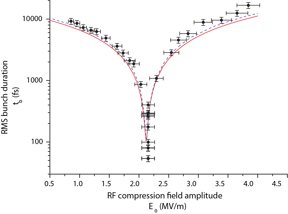
van Oudheusden et al. Phys. Rev. Lett. 105, 264801 (2010)
In 2010 the group at Eindhoven University of Technology demonstrated the compression of 95 keV, space-charge-dominated electron bunches to sub-100 fs durations. These bunches have sufficient charge (200 fC) and are of sufficient quality to capture a diffraction pattern with a single shot, as was demonstrated in a transmission diffraction experiment on a polycrystalline gold foil. Compression is realized by means of velocity bunching by inverting the positive space-charge-induced velocity chirp. This inversion is induced by the oscillatory longitudinal electric field of a 3 GHz radio-frequency cavity. The figure shows the pulse length at the position of the sample for different compression field amplitudes. 100-fold compression of 0.25 pC electron bunches to sub-100 fs root-mean square (rms) bunch durations is achieved.
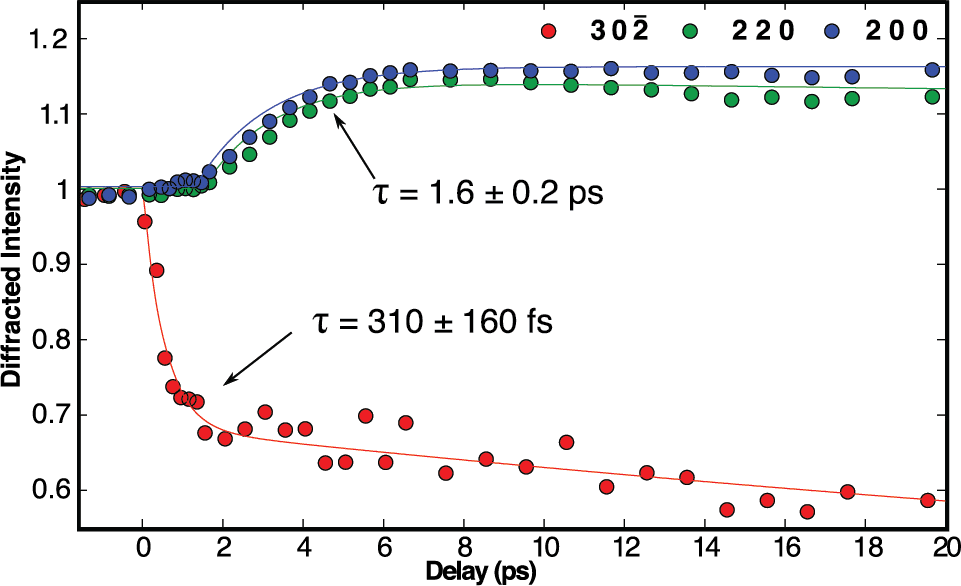
Phase transitions in VO₂
Morrison et al. Science 346, 445-448 (2014)
The complex interplay among several active degrees of freedom (charge, lattice, orbital, and spin) is thought to determine the electronic properties of many oxides. This featured article reports on combined ultrafast electron diffraction and infrared transmissivity experiments in which the authors directly monitored and separated the lattice and charge density reorganizations that are associated with the optically induced semiconductor metal transition in vanadium dioxide (VO₂). By photoexciting the monoclinic semiconducting phase, the authors were able to induce a transition to a metastable state that retained the periodic lattice distortion characteristic of the semiconductor but also acquired metal-like mid-infrared optical properties. The authors demonstrate that ultrafast electron diffraction is capable of following details of both lattice and electronic structural dynamics on the ultrafast time scale.